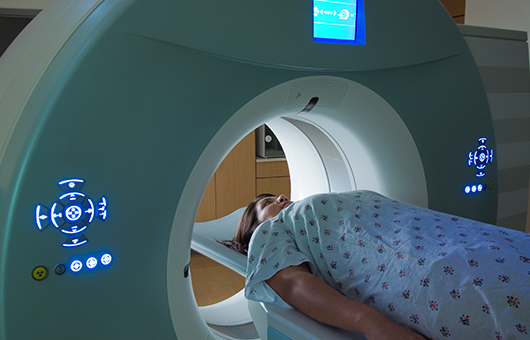
Positron Emission Tomography (PET)
What Is Positron Emission Tomography (PET) ?
Positron Emission Tomography, commonly known as PET, is a powerful medical imaging technique that provides detailed information about the metabolic and biochemical processes within the body. It involves the use of a radiopharmaceutical, which emits positrons, a type of subatomic particle. PET scans are particularly useful for diagnosing and monitoring various medical conditions, including cancer, neurological disorders, and heart disease. PET imaging is performed in specialized facilities by trained healthcare professionals, and strict safety measures are followed to ensure patient well-being. It is a valuable tool in modern medicine for diagnosing and managing a wide range of medical conditions.
- Radiopharmaceutical Injection: A small amount of a radiopharmaceutical is administered to the patient, usually by injection. This radiopharmaceutical contains a radioactive isotope, such as fluorine-18, that emits positrons.
- Positron Emission: Inside the body, the positrons emitted by the radiopharmaceutical interact with electrons, resulting in the release of gamma rays. These gamma rays are emitted in opposite directions, which is a characteristic feature of positron annihilation.
- Gamma Ray Detection: The gamma rays are detected by a ring of detectors surrounding the patient. The detectors measure the location and intensity of the gamma ray emissions.

What Is The Main Cause Of Positron Emission Tomography (PET) ?
The main cause of Positron Emission Tomography (PET) is the use of a radiopharmaceutical, which contains a radioactive isotope that emits positrons, a type of subatomic particle. This radiopharmaceutical is administered to the patient, typically by injection.
- Radiopharmaceutical Administration: A small amount of a radiopharmaceutical is introduced into the patient's body, usually by injection into the bloodstream. This radiopharmaceutical contains a radioactive isotope, such as fluorine-18, which emits positrons.
- Positron Emission: Inside the body, the radioactive isotope undergoes a process called positron emission. Positrons are positively charged particles, which are the antimatter counterpart of electrons.
- Positron Annihilation: The emitted positrons encounter electrons within the body. When a positron collides with an electron, both particles are annihilated, and their mass is converted into energy in the form of two gamma rays traveling in opposite directions.
- Gamma Ray Detection: The gamma rays produced during positron annihilation are detected by a ring of specialized detectors surrounding the patient.
- Image Reconstruction: The data collected by the detectors is sent to a computer, which processes the information and generates detailed 3D images of the distribution of the radiopharmaceutical within the body.
What Is Treatment ?
Treatment Positron Emission Tomography (PET) refers to the use of PET imaging in combination with therapeutic interventions, typically for cancer treatment. This approach, known as PET-guided therapy or PET-directed therapy, involves using PET scans to guide and monitor the delivery of targeted treatments. Here's how treatment PET works: Radiopharmaceutical Administration: Similar to diagnostic PET, a radiopharmaceutical is administered to the patient. However, in treatment PET, the radiopharmaceutical may be labeled with a different radioactive isotope that is suitable for both imaging and therapy. Imaging and Dosimetry: The patient undergoes a PET scan to visualize the distribution of the radiopharmaceutical within the body. This information is used to determine the absorbed dose of radiation delivered to the target tissue and surrounding areas. Treatment Planning: Based on the PET images and dosimetry data, a treatment plan is developed. This plan outlines the amount of radiation to be delivered, the specific target areas, and any necessary precautions to protect nearby healthy tissues. Radiation Therapy Delivery: The patient receives radiation therapy using a specialized treatment system. This may involve external beam radiation therapy (where radiation is directed from outside the body) or internal radiation therapy (where radioactive sources are placed inside the body near the tumor). Ongoing Monitoring: Throughout the course of treatment, additional PET scans may be performed to assess the response to therapy and make any necessary adjustments to the treatment plan. Treatment PET is particularly valuable in situations where precise targeting of radiation therapy is crucial. It allows healthcare providers to visualize the distribution of the therapeutic radiopharmaceutical and ensure that the intended treatment area receives the appropriate dose of radiation. One example of treatment PET is the use of radiopharmaceuticals like Lutetium-177 PSMA for advanced prostate cancer. The radioactive substance targets prostate-specific membrane antigen (PSMA), a protein found on the surface of prostate cancer cells. This allows for precise delivery of radiation to the cancer cells while minimizing damage to surrounding tissues. Treatment PET is a specialized approach that requires a multidisciplinary team of healthcare professionals, including nuclear medicine physicians, radiation oncologists, and medical physicists, to plan and administer the therapy effectively.
Clinical Services
Facilities
24 Hours Services


