
Computed Tomography (CT)
What Is Computed Tomography (CT) ?
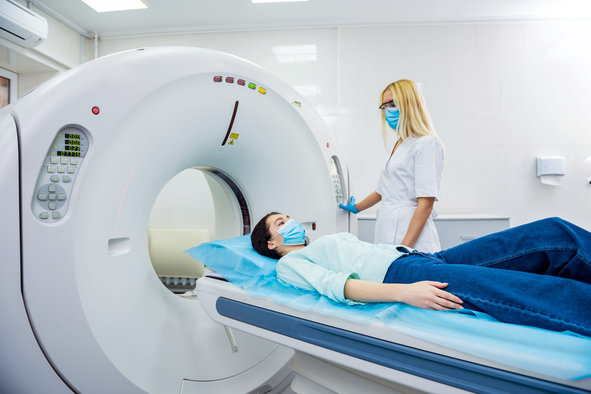
Computed Tomography, commonly known as CT or CAT scan, is a medical imaging technique that uses X-rays and advanced computer processing to create detailed cross-sectional images of the inside of the body. It provides a more comprehensive and three-dimensional view compared to traditional X-ray imaging. CT scans are particularly useful for visualizing soft tissues, blood vessels, organs, and bones. They are commonly used to diagnose a wide range of conditions, including trauma, cancer, infections, and vascular diseases. Additionally, CT scans can be used to guide interventional procedures, such as biopsies or the placement of drainage tubes. It's important to note that CT scans involve exposure to ionizing radiation, and healthcare providers take measures to ensure that the benefits of the scan outweigh any potential risks. The technology and protocols used in CT scans continue to advance, allowing for lower radiation doses while maintaining image quality.
- X-ray Beams: Like conventional X-ray imaging, CT involves the use of X-ray beams. In a CT scanner, an X-ray tube rotates around the patient, emitting a series of narrow X-ray beams.
- Detectors: On the opposite side of the patient, there is a ring of detectors that capture the X-ray beams after they pass through the body. These detectors measure the amount of radiation that reaches them.
- Data Acquisition: As the X-ray tube and detectors rotate, multiple X-ray images are acquired from different angles. This data is then sent to a computer for processing.
- Reconstruction: The computer processes the collected data and uses sophisticated algorithms to reconstruct a series of cross-sectional images, or "slices," of the body. These slices provide detailed views of the internal structures.
- 3D Visualization: The reconstructed images can be viewed in various planes (axial, sagittal, and coronal) and can be manipulated to create 3D visualizations, allowing healthcare professionals to examine the body from different perspectives.
What Is The Main Cause Of Computed Tomography (CT) ?
The main cause of Computed Tomography (CT) is the use of X-ray radiation to create detailed cross-sectional images of the inside of the body. CT scanners combine X-ray technology with advanced computer processing to generate these images.
- X-ray Beams: A CT scanner uses an X-ray tube that emits a series of narrow X-ray beams. These X-ray beams pass through the body and are detected on the other side.
- Rotating X-ray Tube and Detectors: In a CT scan, the X-ray tube and a ring of detectors are mounted on opposite sides of a circular gantry. The gantry rotates around the patient during the scan.
- Data Acquisition: As the gantry rotates, the X-ray tube emits X-ray beams from multiple angles. The detectors measure the amount of radiation that passes through the body and reach them. This data is collected and sent to a computer.
- Reconstruction: The computer processes the collected data and uses mathematical algorithms to reconstruct a series of cross-sectional images, or "slices," of the body. These slices provide detailed views of internal structures.
- Image Visualization: The resulting images can be viewed on a computer monitor and can be manipulated to view different planes (axial, sagittal, and coronal) or to create 3D visualizations.
What Is Treatment ?
Treatment Computed Tomography (CT) refers to the use of CT scanning technology in the context of radiation therapy, a form of cancer treatment. In this application, CT scans are used to precisely plan and deliver radiation therapy to target and treat cancerous tumors while minimizing damage to surrounding healthy tissues. Treatment CT is an integral part of modern radiation therapy, allowing healthcare professionals to plan and administer treatment with a high degree of accuracy. By using CT images for treatment planning and guidance, radiation oncologists can tailor the therapy to the individual patient's anatomy and the specific characteristics of their cancer, maximizing the effectiveness of the treatment while minimizing potential side effects to surrounding healthy tissues.
Clinical Services
Facilities
24 Hours Services
@ 2023 Utkarsh Hospital kota